Day 1 :
Keynote Forum
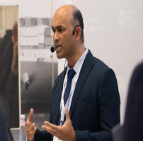
James C.M. Hwang
Professor & IEEE - Distinguished Microwave Lecturer, Cornell University, USA
Keynote: Microwaving a Biological Cell Alive ‒ Label-free Noninvasive Cell Characterization by Broadband Impedance Spectroscopy

Biography:
James Hwang is Professor in the Department of Materials Science and Engineering at Cornell University. He graduated from the same department with a Ph.D. degree. After years of industrial experience at IBM, Bell Labs, GE, and GAIN, he spent most of his academic career at Lehigh University. He co-founded GAIN and QED; the latter became the public company IQE. Between 2011 and 2013, he was the Program Officer for GHz-THz Electronics at the U.S. Air Force Office of Scientific Research. He has been a visiting professor at Cornell University in the US, Marche Polytechnic University in Italy, Nanyang Technological University in Singapore, National Chiao Tung University in Taiwan, Shanghai Jiao Tong University, East China Normal University, and University of Science and Technology in China. He is an IEEE Life Fellow and a Distinguished Microwave Lecturer. He is also a Track Editor for the IEEE Transactions on Microwave Theory and Techniques. He has published more than 350 refereed technical papers and been granted eight U.S. patents. He has researched for decades on the design, modeling and characterization of optical, electronic, and micro- electromechanical devices and circuits. His current research interest focuses on electromagnetic sensors for individual biological cells, scanning microwave microscopy, and two-dimensional atomic-layered materials and devices.
Abstract:
Microwave is not just for cooking, smart cars, or mobile phones. We can take advantage of the wide electromagnetic spectrum to do wonderful things that are more vital to our lives. For example, microwave ablation of cancer tumor is already in wide use, and microwave remote monitoring of vital signs is becoming more important as the population ages. This talk will focus on a biomedical use of microwave at the single-cell level. At low power, microwave can readily penetrate a cell membrane to interrogate what is inside a cell, without cooking it or otherwise hurting it. It is currently the fastest, most compact, and least costly way to tell whether a cell is alive or dead. On the other hand, at higher power but lower frequency, the electromagnetic signal can interact strongly with the cell membrane to drill temporary holes of nanometer size. The nanopores allow drugs to diffuse into the cell and, based on the reaction of the cell, individualized medicine can be developed and drug development can be sped up in general. Conversely, the nanopores allow strands of DNA molecules to be pulled out of the cell without killing it, which can speed up genetic engineering. Lastly, by changing both the power and frequency of the signal, we can have either positive or negative dielectrophoresis effects, which we have used to coerce a live cell to the examination table of Dr. Microwave, then usher it out after examination. These interesting uses of microwave and the resulted fundamental knowledge about biological cells will be explored in the talk.
Keynote Forum
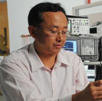
Sandeep K. Vashist
Global IVD Director, Pictor Diagnostics Pvt. Ltd, Germany
Keynote: TBA

Biography:
Dr. Sandeep K. Vashist is the Global IVD Director, Pictor Diagnostics Pvt. Ltd in Germany. After completing his Ph.D. from Central Scientific Instruments Organization, India, in 2006, he was the Bioanalytical Scientist at Bristol-Myers Squibb Ireland (2006−2009), the Team Leader for point-of-care (POC) diabetic monitoring at NUSNNI, Singapore (2009−2012), and the Head of Immunodiagnostics at Hahn-Schickard, Freiburg, Germany. His research outputs include many successful technology transfers; several in vitro diagnostic (IVD) and POC technologies; more than 100 journal publications, 6 patents, 8 book chapters, more than 90 conference publications, many invited/keynote/plenary talks, and 3 forthcoming books. He has constantly received highly prestigious fellowships and awards for scientific research excellence and innovation. He is the Executive Editor, Editorial Board Member and Guest Editor for >10 high impact factor journals (Sci Rep, BB Reports, NPBR, Diagnostics, Sensors, and others). Besides, he is the expert/reviewer/advisor for many international funding agencies, and a consultant for IVD, POCT and diabetic monitoring and management.
Abstract:
TBA
- Oral Presentation
Session Introduction
Dugan Um
Texas A&M University – Corpus Christi, USA
Title: Microscale surface reconstruction by structure from motion for biological applications
Time :

Biography:
Dugan Um achieved his PhD in the study of sensitive robotic skin for unknown environments motion planning. After he received his degree, he joined Caterpillar Inc. as a research engineer and worked for about 4 years at Caterpillar R&D group and Research center. Currently he is teaching at Texas A&M University, Corpus Christi delivering his 4 years of engineering experiences into classes. His research areas include robotic motion planning, 3D sensing, Reinforce learning, and MEMS technology. He is currently an associate professor of Mechanical Engineering and Geospatial Computing Sciences, Texas A&M University at Corpus Christi, USA.
Abstract:
In this paper, we demonstrate a novel microscale surface reconstruction technology by Structure from Motion (SfM) for biological applications. Demands in 3D surface reconstruction of microscale parts for biological applications is ever increasing for molecular design, microscale geno/phenotyping, etc. In microscale photogrammetry, the confocal microscopic imaging technique has been the dominant trend. We propose a novel method to construct a 3D shape in microscale with less size limit of an object. Recently, the surface from motion (SfM) demonstrated reliable 3D reconstruction for macroscale objects. In this paper, we discuss the results of a novel micro-surface reconstruction method using the Surface from Motion in microscale. The proposed Micro SfM technique utilizes the photometric stereovision via microscopic photogrammetry. The main challenges lies in the scanning methodology, ambient light control, and light conditioning for microscale objects. Experiments with light sensitive aspects of the SfM in microscale has been shared and will be addressed in the paper.
- Young Research Forum (Oral)
Session Introduction
Travis McCain
Flux Biosciences, Inc. San Francisco, USA
Title: Comparison of urinary hormone measurements between novel giant magnetoresistive (GMR) biosensor and conventional enzyme immunoassay (EIA)

Biography:
Travis W. McCain, MS is Chief Product Officer of Flux Biosciences, Inc. USA. In June, 2021 he will confirm both MD and MBA degrees from Dartmouth College, USA. He holds an MSc in Cellular and Integrative Physiology from Indiana University, USA and BSc in Engineering, Product Design from Stanford University, USA.
Abstract:
Introduction: Ovulatory disorders, such as Polycystic Ovarian Syndrome are a common cause of infertility in women. The hormones relevant to ovulatory disorder diagnosis are present in healthy women and the concentration of these hormones in urine varies throughout the menstrual cycle, over the course of a woman’s life, and are affected by a variety of other lifestyle and medical factors. Objective: Characterize the performance of a portable giant magnetoresistive (GMR) biosensor in measuring the concentration of several urinary hormones related to ovulatory disorders in women. Participants: 5 women participated over the course of 28 days. Methods: Participants provided a brief medical and menstural history upon enrollement into the study. Over the course of the study, the concentrations of lutenizing hormone (LH) and pregnanediol-3-glucuronide (PDG) in first morning urine were measured by prototype GMR platforms and commercially available EIA. Urinalysis, creatinine measurements and biotin concentrations were also gathered on each sample. Results: forethcoming. Conclusions: GMR biosensor offers a promising method for quantitative measurement of LH in point of care format. Further assay development is indicated, especially for PDG. Biotin in urine, presumably from supplementattion appears to affect performance of GMR assay utilizing biotinylated antibodies.
Geraldine Isamari Silva Galindo
PhD Student, Research Center in Applied Science and Advanced Technology (CICATA), Mexico
Title: The role of titanium dioxide (TiO2) in the construction of nonenzymatic glucose biosensors

Biography:
Geraldine Isamari Silva Galindo has complete her university at 23 years from Instituto Politécnico Nacional, in bionic engineering; at the present time, she is studying a master in advanced nanotechnology, and she has been working in the development of nonenzymatic and noninvasive glucose biosensors, based on oxides as TiO2, CuO, and NiO, synthesized by physical techniques as RF-sputtering reactive and electrochemical techniques, and TiO2 and carbon nanotubes synthesized by chemical techniques.
Abstract:
In recent decades, the importance of catalytic processes in various areas of application has brought about the optimization of various materials to provide systems with higher reaction rates, especially in the construction of nonenzymatic glucose biosensors, due to the high demand of continuous glucose monitoring; currently, modern glucose biosensors are based on heterostructures of various materials. It has been demonstrated that it is possible to improve catalysis processes by adding supports, controlling their physical and chemical synthesis properties. The objective is that the main catalysis process must be carried out on a support that facilitates the transfer of electrons between the catalytic material and itself, providing stability to the particles that carry out the catalysis processes, and promoting the performance of catalytic materials, thus reducing costs in fabrication, since most of them are high-cost noble metals such as gold and platinum. TiO2 has demonstrated to be a great material for the construction of the supports due to its physical and chemical stability in an alkaline and acid environment and its low cost of mass production. In this work is presented the methodology to synthesize TiO2 anatase supports electrodes by Pechini method and its morphological and structural characterization; it is also presented the electrochemical characterization which includes the behavior of the electrodes at different pH conditions, the study of selectivity and sensitivity in the presence of glucose concentrations and possible interferents in non-invasive fluids as urine, and the reproducibility and stability of the electrodes by techniques as cyclic voltammetry and chronoamperometry.
Sebastian Horstmann
PhD Student, University of Cambridge, UK
Title: A capacitive touchscreen biosensor for label-free detection of electrolyte concentrations

Biography:
Sebastian Horstmann received his BSc and MSc degree in Medical Physics from the Heinrich Heine University of Duesseldorf. He started his research on colloidal particles in laser potential fields in Prof Stefan Egelhaaf’s Experimental Soft Matter Physics Group in Duesseldorf and focused on droplet microfluidics as a visiting graduate student in Prof David Weitz’ Experimental Soft Condensed Matter Laboratory at Harvard University. He holds an MRes degree in Sensor Technologies and Applications and is currently pursuing an EPSRC funded PhD on capacitive touch sensing of electrolytes and biomaterials for healthcare applications in Dr Ronan Daly’s Fluids in Advanced Manufacturing Group at the Institute for Manufacturing and in Prof Lisa Hall’s Analytical Biotechnology Laboratory at the University of Cambridge.
Abstract:
We are investigating the use of mutual projected capacitive touchscreens as a label-free sensing technique. It is projected that by 2021 the number of users of capacitive touchscreen-based mobile devices will have grown to 3.8 billion people worldwide, a 52% increase within just five years. Smartphone penetration is especially expanding amongst developing countries and has attracted attention for collecting a sensor’s readout or interpreting sensed data in low-cost healthcare applications. In the medical technology sector, in-vitro diagnostics is the leading growth area and anticipated applications include biosensors will allow monitoring of drinking water quality or measurement of physiological parameters such as blood glucose levels. Here we report on the benefits and challenges of using the capacitive touchscreen for biosensing studies, a component of the mobile device often neglected in the literature. Capacitive fringe fields project above the touchscreen’s glass surface to sense and interact with a stylus or finger. Instead, we examine interactions with fluid samples and specifically sensing the presence of electrolytes. This is carried out by studying the polarisation properties of electrolyte samples in response to different electrical perturbation frequencies up to the megahertz regime. Initial results show a linear response for static capacitance measurements of low ionic concentrations below 200 µM of sodium, magnesium, calcium or potassium chloride. This has potential to directly transfer to human sweat sensing for monitoring of chronic kidney dysfunction and opens the door to exploring more complex biosensing with capacitive touchscreen displays.
Ishita Auddy
PhD Student, Indian Institute of Food Processing Technology, India
Title: Development of amperometric multi-enzyme biosensor to evaluate the adulteration in Virgin coconut oil (VCO)

Biography:
Ishita Auddy pursuing a PhD from Indian Institute of Food Processing Technology (IIFPT), India and at the age of 26years completed her Master's Degree.
Abstract:
Virgin coconut oil (VCO) is costly, and due to this reason, the marketers opt for adulteration with coconut oil. Diglyceride is the only parameter in VCO which can be used to identify adulteration with coconut oil. The existing analytical techniques used to detect the diglyceride content are based on chromatographic techniques, Nuclear Magnetic Resonance (NMR) and other spectroscopic methods. However, these methods are complicated, time-consuming. Hence, an enzyme-based amperometric biosensor may be the feasible solution to determine the diglyceride content in VCO. A multi enzyme-based amperometric biosensor was developed using a three-electrode type system. The characterization study of the immobilized working electrode was done using a Scanning Electron Microscope (SEM) and cyclic voltammetry. The working electrode process parameter like potential, % glutaraldehyde, gelatin concentration, BSA concentration and pH of the buffer has been optimized. The empirical relationship was developed between the diglyceride and output current. Different ratios of adulterated samples were taken, and the validation of the biosensor was done using HPLC method. The maximum response was found at the potential of -0.4V with 2.5%gluteraldehyde and 45 mg of gelatin at pH 7.0. Linear empirical relation developed was found to have a coefficient of determination (R2) =0.99. The validation study showed no significant difference at 95 % confidence level. The precision (CV%) was found to be 0.15±0.56 and 0.1±0.32 for the first day and seventh day, respectively. The detection limit was found in the concentration range from 2ppm to 1200 ppm of diglyceride with a detection time of 10 seconds per samples. Hence, the developed biosensor can be used to evaluate the adulteration in VCO and was observed to be reused for about 15 days.
Rhea Patel
PhD Student, Indian Institute of Technology Bombay, India
Title: D Detect: Development of µTAS vitamin D detection technique

Biography:
Rhea Patel has completed MSc in Biochemistry and currently pursuing PhD from IIT Bombay, India in the field of Biosensors. She has experience in the field of Lab on Chip technology, nanotechnology and has about 4 publications in the same.
Komal Joshi has completed MSc in Biotechnology and currently pursuing PhD from IIT Bombay, India in the field of cancer immunotherapy. She has experience in the field of nanotechnology and published more than 10 publications in the same.
Vishnuram Abhinav is a competent PhD student in the Electrical Engineering Department of Indian Institute of Technology Bombay, India having experience in the development of Lab-on-Chip devices for healthcare, and has five publications.
Parijat Deshpande is a TCS Senior Scientist and pursuing - PhD at the Indian Institute of Technology, Bombay, India, and working on Novel Wearable Sensing Apps.
Abstract:
Changing lifestyles has rendered human beings more prone to dietary deficiencies. One of such prevalent deficiencies is vitamin D, a vitamin extremely important for the bone homeostasis. The deficit of vitamin D has been observed in individuals ranging over all age groups however, the population under threat involves, infants, pregnant women, women in post-menopausal age, old age people, and individuals suffering from autoimmune bone related disorders. Thus, the detection of vitamin D has become the 5th most popular testing in diagnostics accounting for approximately $ 605.9 million worldwide in the year of 2018.
Currently, vitamin D testing is being done by the use of techniques that involve sophisticated instrumentation and skilled labor thus restricting the feasibility of testing to modernized diagnostic laboratories in urban areas. In order to make the testing available to the population in the developing countries, we have designed a cost-effective and user-friendly solution in the form of an enzyme coupled impedance based portable sensor for the detection of vitamin D.
The device is based on the in-vivo activation process of vitamin D that takes place via oxidation of 25-dihydroxyvitamin D3 to 1α,25-dihydroxyvitamin D3 [1α,25(OH)2D3] by enzyme CYP27B1. The D detect involves electrode immobilized with CYP27B1 that reacts with vitamin D in the blood sample and the interaction leads to oxidation of vitamin D. The interaction leads to a change in impedance that is being amplified and detected with the help of portable potentiostat. The D detect serves as a simple and cost effective point of care diagnostic for vitamin D.
Ganesan Sivaramakrishnan
PhD Student, University of Lille, France
Title: Highly sensitive smart biosensor based on the surface plasmon resonance (SPR)

Biography:
Ganesan Sivaramakrishnan is a third-year PhD student from the University of Lille, France. His PhD will be finishing in September 2020. He published an article related to super-resolution microscopy in the nature communication journal and submitted two articles in the field of biosensing towards a point of care in a reputed journal (will be published soon) and finally writing another article in the field of biosensing which will be soon submitted.
Abstract:
In the current technologic era biosensors are being used largely in different fields of application. Current research trends focus on the invention of a portable detection system with high sensitivity, accuracy, repeatability, and other criteria relevant to the applications. From all the existing biosensors, Surface plasmon resonance (SPR) sensing technology has received continuous attention due to its advantage of a high-sensitivity, label-free, and fast response time. Although the SPR sensing technique being a legend in the sensor community, currently the temperature of the sample needs to be carefully maintained and controlled because the SPR signal varies with temperature. This gives a huge challenge in the application whether the SPR sensor senses only the biological reaction or it detects also the temperature change in the sample. Taking all the challenges into account we have developed a novel SPR sensor design having multi SPR channels allowing to modulate the temperature of each channel preciously in real-time independent to the other channels by using joules effect. We have experimentally demonstrated that the temperature modulation of the SPR channel by Joules heating is reliable and enables the SPR sensor system to still being portable and user-friendly.
Jean Pierre Ndabakuranye
PhD Student, University of Melbourne, Australia
Title: Feasibility of self-monitoring of blood bilirubin levels by dual wavelength approac

Biography:
Jean Pierre Ndabakuranye is a PhD student at the University of Melbourne, Australia from 2017. He received his BSc degree from the National University of Rwanda (2010-2013) and his MSc from Hallym University, South Korea (2014-2016). His research interests are in optical MEMS, bioelectronics, and biomedical engineering technology.
Abstract:
Bilirubin is clinically confirmed as a biomarker for liver health and has been utilized for assessing the prognosis of cirrhosis. Optical and chemical methods have been utilized to determine bilirubin levels in the blood. Whilst optical methods offer real-time monitoring, are handy and immune to infection among other substantial benefits, measurements may not be practical in some instances owing to the instrument complexity and space requirements. In this paper, we investigate the feasibility of using the dual wavelength technique with the aim of miniaturizing the setup. Our experiments were performed using blood phantoms within the pathological range projected from a healthy person to a cirrhotic patient (0.15 - 30 mg/dL). Results show a high sensitivity of bilirubin in blood phantoms with a larger predictive accuracy at higher bilirubin concentrations. This may provide an alternative means of determining blood bilirubin levels mostly for cirrhotic patients.

Biography:
Rabia Asghar is doing her PhD from Beijing Institute of Technology, Beijing, China. She is working on homogenous/heterogenous fluorescent assays. Also opening the conceptual folds of these studies with the comparitive studies on comercial applications of these assays. Development of colorimetric based senor for clinical applications is also included in her on going projects. She also worked on preapring materials (Graphene Oxide, Mollybednum disulphide) for the development of clinically reliable senosrs to detect protein bsed disease biomarkers.
Abstract:
FRET-based sensors turn on /off on the target recognition is a rapid, sensitive and specific technique to detect different targets. Graphene and its composite for the quenching of fluorescent in FRET-based homogenous assays have been reported in many studies. Graphene/graphene composites are efficient energy transducers in FRET-based biosensors and repeatedly used in many studies up till now. In many fluorescent assays, these are used to quench the fluorescence of labelled single-stranded DNA when these labelled strands (DNA/RNA) are attached to the surface of quenchers. Upon getting high affinity and specific target the quenched labelled strands detached from the quencher to get bind to the relevant target. Here in this study, we labelled both ends of the aptamer with the fluorophores of two different wavelengths and observed under fluorescence microscope. To study quenching and restoration of fluorescence on a highly specific end of the aptamer. The whole study observed under the fluorescence microscope. Fluorescence –quenching – the restoration of fluorescence is followed by attachment of aptamer onto the quenchers surface and restoration of fluorescence when it detaches from rGO to attach with the target. Graphene oxide prepared by a hydrothermal process in our laboratory. To enhance energy transduction we reduced the graphene oxide surface. In this study, we proposed the binding of aptamer onto the surface of quencher is not all due to π-bond stacking but involved electrostatic attractions more dominantly. Reduced surface allows the restoration of fluorescence more efficiently. We used thrombin because of the high specification to its 15-mer DNA oligonucleotideMoreover, we also open the clinical aspects of such studies and their reliability to use these assays in clinical, industrial and laboratory applications.


