Day 2 :
Keynote Forum
Andreas Dietzel
Center for Pharmaceutical Engineering (PVZ), Germany
Keynote: Microfluidic systems for pharma technology - the manipulation of cells, droplets and particles
Time : 09:00-09:40

Biography:
Andreas Dietzel studied Physics and completed his PhD at University of Göttingen in 1990. In the years 1990 to 2003, he worked in different organizations of IBM including the Research Laboratory in Rüschlikon. In 2004, he joined TU Eindhoven as a Full Professor of Micro and Nanoscale Engineering. In 2012, he was appointed as Professor at TU Braunschweig and Director of the Institute of Micro-technology. His research interest focuses on “The design and fabrication of microsystems and especially of microfluidic systems with applications in the life sciences”
Abstract:
In a world that becomes increasingly concerned about affordable health care, fast and effective screening methods for drugs in different formulations are required in the course of their development. In addition, the trend towards personalized medicine demands production of drugs in very small volumes. For both, the microfluidic approach is ideally suited. With miniaturized systems that can be realized by micro- or nanofabrication processes, new tools for pharmaceutical research and development become available. As new and better technologies for pre-clinical screening of drug dosage formulations microfluidic cell culture models that can mimic in-vivo conditions have attracted much attention. Recently developed organ-on-chip platforms providing dynamic flow conditions like cornea-on-chip and pancreas-on-chip will be presented including aspects of their microfluidic design, their fabrication and application. These systems are equipped with integrated sensors but also allow microscopic access at low background auto fluorescence. Furthermore recent work on the production of nanoparticles formulations within microfluidic droplet flows and plug flows will be discussed. In thereby obtained smallest fluid volumes mixing is accelerated and very controlled precipitation occurs. This leads to nanoparticle formulations in which particle sizes can be tuned by external flow controls. These approaches offer new possibilities for production at smallest scales and for improving the bioavailability of poorly soluble drugs.
Keynote Forum
Oliver Hayden
Siemens Healthcare GmbH, Germany
Keynote: Why magnetic sensing can be a useful method for in-vitro cell diagnostics
Time : 10:00-10:40

Biography:
Oliver Hayden is a Scientist who leads in-vitro diagnostics and bioscience research at Siemens Healthcare GmbH. Before joining Siemens, he was a Visiting Scientist at IBM Research, Switzerland, working on Post-CMOS technologies. He performed Post-doctoral research at Harvard University on nano-photonics. He completed his Doctoral degree in Biochemistry working on “Molecularly imprinted polymers”
Abstract:
Blood is the most important source for routine in-vitro diagnostic information, such as the concentration level of plasma biomarkers and cells. Only in special cases, such as leukemia diagnostics, functional analysis is performed with fluorescence flow cytometry. However, the rich information content of blood cell functions is not routinely available due to the complexity of today’s diagnostic workflow. Furthermore, sample logistics and sample preparation efforts cause imprecision of the test results. In my presentation, I review the single cell analysis challenges with opaque whole blood as sample matrix and the clinical unmet need for point of care usability. To demonstrate how cell function diagnostics can be achieved at the point of care, I will discuss my research efforts to integrate microfluidic workflows and giant magnetoresistance sensors.
Keynote Forum
Xiaohao Wang
Tsinghua-Berkeley Shenzhen Institute, China
Keynote: Microfluidic chip ionization source coupling with mass spectrometry
Time : 09:40-10:20

Biography:
Xiaohao Wang completed his Bachelor and Doctor degree both at Tsinghua University in 1994 and 1999, respectively. From 1998, he worked at Tsinghua University as a Faculty member, and was promoted to Full Professor at 2000. From 2007 to 2008, he was at Technical University of Berlin as a Visiting Scholar. He serves as the Deputy Dean of Graduate School at Shenzhen, Tsinghua University, now. His research interests cover MEMS based sensors and actuators, ionizing sources and portable analytical instrument. He has published over 200 technical papers and holds tens of patents.
Abstract:
Ionization source is a vital component of the mass spectrometer. In recent years, with the development of the mass spectrometer miniaturization and the microfluidic chip integration technologies, increasing research efforts have been devoted to the coupling of microfluidic chip ionization source to mass spectrometry. Facing requirement of portable MS used for on-site rapid detection, a microfluidic chip ionization source is developed. Multi-layer soft photolithography technology is chosen as the fabrication craft for the microfluidic chip template, and three novel microfluidic chip ionization sources were proposed, such as a microfluidic chip-based multi-channel ionization (MCMCI) was developed to extend the application of microfluidic chip ionization to MS. This MCMCI implemented extraction of untreated compounds in complex matrices without sample pretreatments and dual sprays with high DC voltages simultaneously.
- Nanotechnology in Biosensors
Biosensor Applications
Biosensing Technologies
Biochips & Nucleic Acid Sensors
Bioinstrumentation & Equipments
Bio-MEMS/NEMS
Photonic Sensor Technologies

Chair
Andreas Dietzel
Center for Pharmaceutical Engineering(PVZ), Germany

Co-Chair
Sarmiza Elena Stanca
Leibniz Institute of Photonic Technology , Germany
Session Introduction
Ike Chi
California Institute of Technology, USA
Title: Recent and upcoming potential spacecraft missions requiring biosensor technologies: Current examples, what are we looking for and remaining challenges
Time : 10:20-10:50

Biography:
Ike Chi is a Materials and Processing Engineer at NASA’s Jet Propulsion Laboratory. He is the integrated product team (IPT) lead for Skutterudite Technology Maturation (STM) program and the device development task lead for Advanced Thermoelectric Couples (ATEC) program. He is also currently a member of Multi-Mission Radioisotope Thermoelectric Generator (MMRTG) Pyroshock project. He received his PhD from the Johns Hopkins University in 2014. He had several years of experience in fabricating biocompatible ceramics/semiconductors and ultra-high surface area materials. He is also interested in the area of biomedical implants/scaffolds and biosensing.
Abstract:
The National Aeronautics and Space Administration (NASA) has upcoming spacecraft missions to Mars (i.e., Mars 2020) and future potential missions (e.g., landers, penetrators) in the planning stages to Mars, Europa, Enceladus and Titan that could require unique biosensor systems to search for critical biomarkers in those environments. In-situ sensing capability under extreme environmental conditions is particularly critical for these current and potential NASA space exploration missions. JPL/NASA’s future planned Europa Clipper multiple flyby mission and a potential Europa lander or the planned Mars 2020 (ESA ExoMars mission) will encounter extreme environmental conditions. This presentation will report on our to-date accomplishments at the Jet Propulsion Laboratory (JPL) on Mars and potential plans in these other extreme deep space environments. Those missions will need ultra-sensitive sensors capable of reliable operation across a very wide range of temperatures. The applications of the highly sensitive sensor developed can include habitat health monitoring for a space station and/or for life detection on an Earth-like planet. In order to help fulfill scientific needs, we have developed a portable and low power in-situ biosensor to detect amino acids using an electrochemical spectroscopy technique. We have also enhanced chemical sensitivity of the sensor to parts-per-billion (ppb) range by integrating novel nanostructured electrode materials with improved surface properties. This novel engineered nanostructured micro-device tailored to sense specific analytes (e.g., amino acids) could be integrated with multiple flight-proven sensing platforms for a wide range of missions. This presentation will report on the progress for validating performance of this multi-platform in-situ bio-sensing device developed and tested by JPL.
Sarmiza Elena Stanca
Leibniz Institute of Photonic Technology, Germany
Title: Secure accuracy at increase precision of AFM-probe integrated biosensor
Time : 11:10-11:40

Biography:
Sarmiza Elena Stanca has her expertise in electrochemical and optical nanosensors achieved during her research activity at the EPFL Lausanne (Swiss Confederation Fellow), UCD Dublin (Marie-Curie-Fellow), UKJ Jena (Marie-Curie-Fellow), University Babes-Bolyai Cluj-Napoca, Research Centre Karlsruhe and IPHT Jena (DAAD Fellow). She is currently a Scientist at the Leibniz Institute of Photonic Technology, Jena.
Abstract:
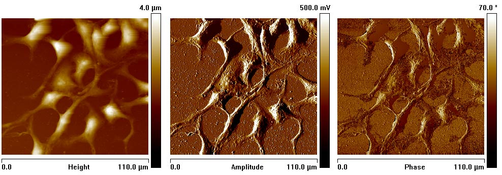
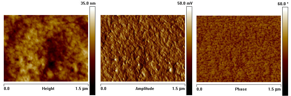
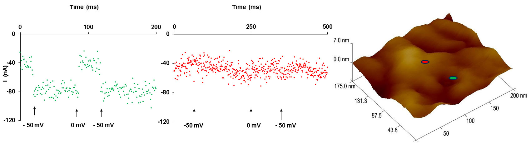
The plasma membrane regulates the selective interchange of matter between the interior and the exterior of the cell. Understanding this complex process requires knowledge of the plasma membrane´s molecular constituents. Topical reports prove the access to the molecular level of the synthetic membrane by atomic force microscopy (AFM). This technique also permits an electrochemical investigation in the immediate vicinity of the tip. An electrochemical and topographic study of the living cell membrane, by the mean of an AFM-probe integrated amperometric biosensors is employed to localize specific molecules in the natural cellular membrane (Figure 1). Several materials and shapes of the AFM probes integrated in different systems are presented. It is underlined that the selection of control experiment is decisive in achieving accurate findings. The central concern of this study is how to preserve the sensor response accuracy while increasing its precision.
Figure 1: (A) Height, amplitude and phase atomic force micrographs (110 µm x 110 µm) of the cells immobilized on conductive glass; (B) Height, amplitude and phase AFMs in one location of 1.5 µm x 1.5 µm of the plasma membrane; (C) AFM probe integrated sensor signal on two different points: green and red marked on the AFM image (200 nm x 200 nm).
A)
B)
C)
Ramla Gary
University of Calabria, Italy
Title: Detection of gold nanoparticles aggregation growth induced by nucleic acid through laser scanning confocal microscopy
Time : 11:40-12:10

Biography:
Ramla Gary has completed her PhD in 2017 from the Laboratory of Liquid Crystals and Interfaces in Physics department in collaboration with the Biological department at University of Calabria, Italy, and Post-doctoral studies from the same University. She has published more than five papers in reputed journals, and has participated in more than eight international and national conferences.
Abstract:
The gold nanoparticle (GNP) aggregation growth induced by deoxyribonucleic acid (DNA) is studied by laser scanning confocal and environmental scanning electron microscope. As in the investigated case, the direct light scattering analysis is not suitable, we observe the behavior of the fluorescence produced by a dye and we detect the aggregation by the shift and the broadening of the fluorescence peak. Results of laser scanning confocal microscopy images and the fluorescence emission spectra from lambda scan mode suggest, in fact, that the intruding of the hydrophobic moiety of the probe within the cationic surfactants bilayer ï¬lm coating GNPs results in a Förster resonance energy transfer. The environmental scanning electron microscopy images show that DNA molecules act as template to assemble GNPs into three-dimensional structures which are reminiscent of the DNA helix. This study is useful to design better nano-biotechnological devices using GNPs and DNA.
Siddheswar Maikap
Chang Gung University, Taiwan
Title: Detection of pH/H2O2 and prostrate/breast cancer biomarker by using nickel-oxide/iridium-oxide sensing membrane in electrolyte-insulator-semiconductor structure
Time : 12:10-12:40

Biography:
Siddheswar Maikap has completed PhD in Department of Physics and Meteorology at IIT Kharagpur on February, 2003. He is Professor at Chang Gung University, Taiwan, since August 2014. He is the holder of three US patents on memory/bio-sensor, eight US/Taiwan patent files, and has more than 100 SCI journal papers, more than 150 international conference papers, 26 keynote/invited talks, and four best paper awards. His recent research focuses on cross-point resistive switching memory for high-density memory as well as bio-sensor applications
Abstract:
Quantification of pH/H2O2 attracts a lot of attentions due to its importance in chemical industries as well as biomedical diagnostic. For the detection of pH and H2O2 by using electrolyte-insulator-semiconductor (EIS) is preferred due to label-free detection, easy fabrication process, and low cost. The NiOx based sensor has shown good pH sensitivity of 50.25 mV/pH. X-ray photo-electron spectroscopy of Ni 2p3/2 has shown two different oxidation states of NiOx membrane and those are Ni2+ and Ni3+ having binding energy 854.5 eV and 856.5 eV, respectively. Existence of these two oxidation states resembles the reduction-oxidation (redox) characteristics of NiOx membrane toward the electroactive species like H2O2. A reference voltage shift of 41 mV is obtained for H2O2 concentration of 10 µM and has shown good linearity up to 100 µM for the first time. In addition, the IrOx membrane shows a record pH sensitivity of 150.4 mV/pH for the first time. This IrOx sensor demonstrated good catalytic behavior as well as the breast cancer biomarker LOXL2 with a concentration of approximately 150 nM is detected. This IrOx nano-net sensor demonstrates good catalytic behavior for H2O2 reduction with a concentration of 100 fM because the oxidation state changes from Ir3+ to Ir4+, whereas a pure SiO2 membrane could not sense H2O2. The oxidation states are confirmed by X-ray photo-electron spectroscopy (XPS). Similarly, prostate cancer is also detected by using NiOx membrane. Therefore, good pH response and redox characteristics of the IrOx/NiOx sensing membrane allow it to diagnosis human disease in future.
Slobodan J Petricevic
University of Belgrade, Serbia
Title: Bi12GeO20 Faraday crystal application in magnetic field measurement

Biography:
Slobodan J Petricevic completed his BSc in Electrical Engineering (EE) in 1996; MSc in EE in 2001 and; PhD in EE in 2007 at School of Electrical Engineering, University of Belgrade, Serbia. His field of research is Optoelectronic and Fiber Optic Instrumentation. He has published 18 scientific papers in SCI listed journal with 105 citations and two patents. He is currently employed as an Associate Professor at School of Electrical Engineering since 2008.
Abstract:
Faraday crystals (FC) have been under intense investigation in magnetic field sensing applications for several decades due to several desirable properties, but mostly due to low interaction with magnetic field that does not disturb the field during measurement. FC requires an optical carrier to sense the magnetic field since interaction of the field and light in the crystal affects the state of polarization of the light. Development of production technology for optical fibers for mass use in telecommunication industry has made design of fiber-optic magnetic field sensor (FOMS) based on Faraday crystal an interesting research field. A class of diamagnetic materials known as sillenites of which BiGeO is an interesting example has been used to sense magnetic field in optical sensor in various configuration and adopted to various application. This paper will discuss an extrinsic, fiber optic, magnetic field sensor, designed for direct point magnetic field measurement constructed using Bi12GeO20 crystal. A configuration suitable for measurement will be presented together with analyses of the test results obtained from a calibrated magnetic field setup. Compensation of temperature effect on magnetic field measurement will be presented and its implication will be discussed.
Umut Kokbas
Cukurova University, Turkey
Title: Detection of β-thalassemia IVSI-110 mutation by using piezoelectric biosensor for non-invasive prenatal diagnosis

Biography:
Umut Kokbas has studied Biotechnology and Biochemistry at Ege University. He is a Research Assistant in Medical Biochemistry department at Cukurova University, working on Thalassemia, which the most common genetic disorder in Turkey. He is also pursuing PhD in the same department.
Abstract:
Statement of the Problem: β-thalassemia is one of the most monogenic autosomal recessive disorders characterized by defective production of the β-chain of hemoglobin. Definition of the β-globin genotype is necessary for genetic counseling in the carriers, and for predicting prognosis and management options in the patients with thalassemia. DNA-based prenatal diagnosis of β-thalassemia routinely relies on polymerase chain reaction (PCR) and gel electrophoresis. The aim of this study is to develop a new procedure, a DNA-based piezoelectric biosensor, for the detection of β-thalassemia IVSI-110 mutation fetuses cell free DNA from maternal blood, the most common β-thalassemia mutation in Turkey.
Methodology & Theoretical Orientation: Cell-free fetal DNA was taken from maternal whole blood. Bioactive layer was constituted by binding 2-hidroxymetacrilate metacriloamidoscystein (HEMA-MAC) nano-polymers on the electrode’s surface. Single oligonucleotide probes specific for IVSI-110 mutation of β-thalassemia were attached to the nano-polymer. The measurements were executed by piezoelectric resonance frequency which is caused by binding of the cell-free fetal DNA in media with single oligonucleotide probe on the electrode surface. The results were confirmed by the conventional molecular method as ARMS.
Findings: The piezoelectric resonance frequencies obtained by hybridization of the cell free fetal DNA on bioactive layer were found 216±12, 273±6, and 321±9 Hz for the samples of normal β-globin, heterozygote, and homozygote of IVSI-110 mutation, respectively.
Conclusion & Significance: The developed biosensor serves as a specific result to IVSI- 110 mutation. It could accurately discriminate between normal and IVSI-110 mutation samples. Because of low costs, fast results, specificity and high detection/information effectiveness as compared with conventional prenatal diagnosis methods, we can offer this technique as an alternative to conventional molecular methods.
Mahamudul Hassan
Murdoch University, Australia
Title: Application of twin-working electrode cell in characterizing biological electron mediators

Biography:
Mahamudul Hassan has completed his BSc (honors) and MS in Microbiology from University of Chittagong, Bangladesh. Currently, he is persuing his PhD at Murdoch Univesity and he aims to investigate the role of electron mediators in microbial extracellular electron transfer (EET) processes.
Abstract:
Electron mediators often play a key role in facilitating microbial extracellular electron transfer (EET) to oxygen or insoluble compounds. This study aims at developing a novel electrochemical cell consisting of two closely (250 µm) mounted working electrodes (WEs), hence Twin-WE; to detect and quantify redox active compounds in a micro-scale (304 µL) environment. A fixed voltage window between two WEs using common counter and reference electrodes was maintained and the individual currents of both WEs were monitored. To detect electron mediators, an optimized voltage window (50 mV) was shifted through a defined potential range (between –1 V and +0.5 V vs. Ag/AgCl) by changing a fixed voltage step (12.5 mV) after the establishment of steady equilibrium current in both WEs. When the voltage window was maintained at the midpoint potential of a mediator, concurrent oxidation and reduction of the mediator occurred as evidence by the concurrent maximal anodic and cathodic current recorded at the two WEs. The electrical current difference plot against the potential scale enabled the identification (by peak location in the potential scale) and quantification (by peak height) of the mediators tested. Our technique enabled a precise determination of riboflavin, anthraquinone-2, 6-disulfonate (AQDS) and two mediators from a pyocyanin producing Pseudomonas aeruginosa (WACC 91) culture both individually and from their mixture. The described Twin-WE cell device is suitable for studying microbial electron transfer processes under a simulated redox environment which prevails in natural habitat. The bio-electrochemical principle underpinning this new method may also be useful for advancing biosensor development.
Stefan Dübel
Technische Universität Braunschweig, Germany
Title: In vitro evolution to adapt antibodies to technical requirements of biosensors

Biography:
Dr. Stefan Dübel is Full Professor of Biotechnology and Director of the respective department at the Technische Universität Braunschweig, Germany. Stefan Dübel serves as a Chair of Biotechnology. He serves as Consultant to biotech and pharma companies. He is initiator of Antibody Factory of the German National Genome Research Network and Editor of the four volumes "Handbook of Therapeutic Antibodies" and other antibody engineering books. He is Co-founder of several biotech companies, most recently of the human antibody provider Yumab GmbH.
Abstract:
The development of biosensors using antibodies for detection of an analyte is frequently hampered by the limited choice of sensor molecules, if antibodies were generated by classical animal immunization. For some antigens or desired fine specificity, no antibodies could be obtained at all. Here, recombinant methods based on a complete in vitro selection pipeline, typically phage display, offer several new opportunities. Antibodies with particular, pre-designed biochemical properties can be selected from the start, as the biochemical milieu during the in vitro selection can be exactly controlled. Further, properties of existing antibodies can be changed or improved in various directions to adapt the sensor molecule to the requirements of a particular biosensor system. We present examples of successful generation of antibodies binding to extremely toxic molecules of antibodies specifically selected to detect very small differences in the antigen structure, matching sandwich pairs, improvement of affinity or stability, and the change of the kinetic binding properties, and successful applications of such recombinant designer antibodies on biosensors.
M W Raad
King Fahd University of Petroleum and Minerals, Saudi Arabia
Title: A home based telehealth system for elderly with Alzheimer’s

Biography:
TBA
Abstract:
By 2020, chronic diseases predicted to account for almost three quarters of all deaths worldwide. According to World Health Organization, the elderly population is expected to become 1.2 billion in 2025. This aging problem contributes greatly to chronic diseases like Alzheimer. The major implications of Alzheimer are patient safety and care. The aim of this paper is to develop a tele-health system, based on internet of things (IoT) technology, for monitoring elderly individuals suffering from Alzheimer’s. We describe a working prototype that is able to capture the vital signs and deliver the desired data remotely for elderly staying at home, using wearable ECG wireless sensor. This prototype has been successfully tested to capture data on a 24/7 basis for a number of patients and volunteer physicians at the KFUPM clinic which helps discover severe cases early on. The developed system includes a suit of signal processing algorithms for the detection of severe cases of arrhythmias in elderly patients. In particular, we use wavelets transform to estimate a number of features from ECG traces which are then used in classification. Our results were benchmarked against the standard MIT physiobank. The performance of the system was also tested on simulated data with very satisfactory results, and very positive feedback from users. In addition, an active wearable RFID wristband, with IR room locators are used to monitor the whereabouts of the elderly at room level.
Elizabeth Salvo
McMaster University, Canada
Title: Towards practical bacterial biosensor assays for on-site applications

Biography:
Elizabeth Salvo is a MSc candidate in Chemical Biology Graduate Program at Biointerfaces Institute-McMaster University working under Dr. John Brennan. She completed her BSc in Chemical Biology at McMaster University.
Abstract:
Air drying in the natural polymers pullulan and acacia gum has been reported previously as an effective bacterial preservation strategy. The use of this method for the preservation of sensing bacteria is presented here. Colorimetric Escherichia coli bioreporters for tetracycline and arsenate analytes has retained responsivity following air-drying in pullulan sugar. Additionally, the method was used to immobilize sensing cells onto paper substrates, and a screen of various materials was conducted to determine the optimal composition of a drying matrix for responsivity. The viscosity enhancer pullulan, acacia gum, polyvinylpyrrolidine and gelatin were paired with osmoprotectants in some standard bacterial culture mediums. The air-drying process is simpler, less expensive, requires less sophisticated instrumentation, and leads to much higher bacterial survival rates compared to the gold standard method for bacterial preservation, lyophilization. Simplicity of the method paired with applicability to simple platforms such as paper test strips makes this research an important step toward the usage of sensing microbes for the detection of a variety of biologically relevant analytes in the field.
M Carmen Bermudo Redondo
Rovira i Virgili University, Spain
Title: Impedimetric electrochemical sensor for albumin determination based on poly bromocresol purple film modified electrodes

Biography:
M Carmen Bermudo Redondo completed her BSc in Chemistry and in Biochemistry at Universitat Rovira i Virgili, Spain and; Post-graduate certificate in Nutrition and Metabolism. She joined the Interfibio-Research Group as a Junior Technician and was rapidly promoted to the position of Senior Technician, actively collaborating in the fields of electrochemical biosensors. She has published nine papers in peer-reviewed journals. In 2016, she started her PhD in Nanoscience, Materials and Chemical Engineering programme at Universitat Rovira i Virgili. Her main research interests include “Nanobiotechnology and molecular biology, ELISA/ELONA techniques, immuno-sensors and apta-sensors, protein extraction, bio-conjugations and novel detection methodologies”.
Abstract:
Human serum albumin (HSA) is the major protein in plasma synthesized principally in the liver and playing significant roles after it is released in the circulation. Albumin is a very important factor of regulation in the exchange of water between the plasma and the interstitial compartment being largely responsible of the colloidal osmotic pressure of blood. Moreover, it is a protein with a notable ability to bind an extensive range of other small molecules helping in the transport of important substances in the human body such as hormones, fatty acids and drugs. A decrease of albumin level in serum is associated with severe illnesses of the kidney as well as other conditions such as liver disease, malnutrition and extensive burns. Therefore, HSA determination is extremely useful in the diagnosis and treatment of many clinical entities. This research work reports on the development and integration of a novel label-free impedimetric sensor based on a poly (bromocresol purple) surface for the specific detection of HSA. The fabricated sensor was incubated with serum albumin, and electrochemical impedance spectroscopy (EIS) was employed to measure the changes in the conductance of the electrode of bromocresol purple (BCP) by reacting with the albumin. In addition, we validated the specificity of the designed sensor to only albumin. Clinical applicability of the sensor was also demonstrated utilizing real serum sample from patients obtaining excellent agreement with the commercial available colorimetric kit. It is expected that the new poly (BCP) sensor will become a successful diagnostic platform for HSA detection in clinical diseases.
Ibragimova Sagila Aladdinovna
State University Dubna, Russia
Title: Synthesis of quantum dots conjugates with antibodies for immunochromatographic analysis

Biography:
Ibragimova Sagila Aladdinovna completed her Graduation with honors at State University of Dubna in Department of Chemistry, New Technologies and Materials and defended her Master's thesis entitled “Synthesizing quantum dot conjugates with monoclonal antibodies to glycoprotein gB of Aujeszky's disease virus for immunochromatographic analysis” in 2016. Her scientific work is related to the immunochromatographic analysis using as markers and quantum dots. She is Co-author of four books, has two patents and participated in nine national and international conferences.
Abstract:
There is large number of different markers used in immunochromatographic analysis (IChA). The use of quantum dots markers fluorescent in near infrared (NIR) region is promising in bioassay. We investigated here sandwich method of IChA. Quantum dots (QDs) exhibit strong and narrow band-edge luminescence. Due to their unique optical properties, QDs are perfect fluorescent markers for molecular diagnostics of diseases. It is possible to synthesize QDs with fluorescence in NIR, where background fluorescence of biological samples is low. Highly bright and photo-stable NIR-emitting CdTeSe/CdS/CdZnS QDs were synthesized following the method. QDs were functionalized with COOH-group using thiol-modified polyvinylpyrrolidone. As a model antigen, we used glycoprotein gB of nonpathogenic for human being Aujeszky's disease virus. QDs were covalently conjugated with monoclonal antibodies to glycoprotein gB of the virus using carbodiimide methods. The conjugates were tested on test strips manufactured by following the method. The investigation showed that the optimal excitation wavelength was of 450 nm, where the background luminescence was low and fluorescence peaks of test and control zones test strip were more pronounced. Calibration curve for the quantitative determination of antigens was drawn in a range up to 25 fmol in the sample. Antigen limit of detection is of 4.2 fmol in the sample.
Nasrin Afsarimanesh
Macquarie University, Australia
Title: MIP-based sensing system for early detection of bone loss

Biography:
Nasrin Afsarimanesh has completed her Bachelor of Engineering in the field of Electronics Engineering in 2006 at Azad University. She then completed her Masters’ degree at Pune University in 2010. Currently, she is pursuing her Doctoral degree at Macquarie University. Her research interest includes “Design and development of smart sensing systems for different bio-medical applications”.
Abstract:
This research proposes a novel real-time, label-free sensing technique for the detection of C-telopeptide of type-I collagen (CTx-I) that can be heelpful in early detection of bone loss. A planar interdigital sensor in conjunction with electrochemical impedance spectroscopy (EIS) was used to study the dielectric properties of the test sample. Molecular imprinted polymer (MIP), including artificial recognition sites for CTx-I molecules was synthesized by precipitation polymerization using CTx-I peptide as a template, ethylene glycol methacrylate as the cross-linker and methacrylic acid as a functional monomer. The sensor was coated using the prepared polymer in order to make the sensor selective for CTx-I molecule. Calibration experiments were performed using different known concentration samples and the reference curve was plotted. Complex non-linear least square (CNLS) curve fitting method was used to estimate electrochemical equivalent circuit parameters. The developed system showed a detection limit of 0.1 ng/ml. Two different unknown samples obtained from sheep blood were measured using the developed sensing system and enzyme-linked immunosorbent assay (ELISA) was used to validate the results.
Biography:
Marlen Zschätzsch has completed her PhD in the year 2013 from VIB, KU Leuven, Belgium. Currently, she is working as a Postdoc at Technical University of Dresden, Germany. Her current research is focused on Biosensor Assay Development
Abstract:
Antibodies have become an increasingly important part of fundamental research and medical applications. To meet the high market demand of antibodies in the biopharmaceutical sector, industrial manufacturing needs to be achieved by large scale, highly productive and consistent production processes. These are subjected to international guidelines and have to be monitored intensely due to high safety standards for medical applications. Surface Plasmon Resonance (SPR) spectroscopy - a fast, real-time and label-free bio-sensing method represents an interesting alternative to the quantification of antibody concentrations by ELISA during antibody production. Towards the application of in-process monitoring of active and total antibody concentrations in cell culture probes, a SPR assay using a target antibody model system was developed. In order to ensure the subsequent detection of active antibody concentrations, suitable immobilization strategies of the target were identified. A significant decrease of the limit of detection (LOD) was achieved by using an adapted Ni-NTA method compared to the commonly used amine coupling. Furthermore, the system showed LOD in the low ng/ mL range similar to control antibody quantifications by ELISA. Moreover, the detection of total antibody concentrations for discrimination of specific antibody production efficiency was shown. In conclusion, the development of an alternative quantification system to monitor antibody production was accomplished by using SPR with the advantage of low analyte volume, short assay time and biosensor reusability by target-layer regeneration. The technical development of a SPR-based overall system for continuous in-process control with Sierra Sensor GmbH using the established method is ongoing.
Oliver Hayden
Siemens Healthcare GmbH, Germany
Title: Why magnetic sensing can be a useful method for in-vitro cell diagnostics

Biography:
Oliver Hayden is a Scientist who leads in-vitro diagnostics and bioscience research at Siemens Healthcare GmbH. Before joining Siemens, he was a Visiting Scientist at IBM Research, Switzerland, working on Post-CMOS technologies. He performed Post-doctoral research at Harvard University on nano-photonics. He completed his Doctoral degree in Biochemistry working on “Molecularly imprinted polymers”.
Abstract:
Blood is the most important source for routine in-vitro diagnostic information, such as the concentration level of plasma biomarkers and cells. Only in special cases, such as leukemia diagnostics, functional analysis is performed with fluorescence flow cytometry. However, the rich information content of blood cell functions is not routinely available due to the complexity of today’s diagnostic workflow. Furthermore, sample logistics and sample preparation efforts cause imprecision of the test results. In my presentation, I review the single cell analysis challenges with opaque whole blood as sample matrix and the clinical unmet need for point of care usability. To demonstrate how cell function diagnostics can be achieved at the point of care, I will discuss my research efforts to integrate microfluidic workflows and giant magnetoresistance sensors.
Jaewon Kim
Sungkyunkwan University, South Korea
Title: Development of miniaturized uniaxial cell stretching device

Biography:
Jaewon Kim has completed his Master’s degree at Sungkyunkwan University. Now, he is pursuing PhD in Mechanobiology at the same university.
Abstract:
Cells react variously to external mechanical stimuli such as shear stress from fluid flow or tensile stress caused by substrate deformation. As a result, their structural and functional properties are largely affected by these mechanical stresses, and this phenomenon has been extensively investigated by using cell stretching devices. However, conventional cell stretching devices still have technical limitations to observe such complicated cellular responses. For example, many cell stretching devices are too large to be placed on the stage of microscope and often generate excessive heat and vibration, which are harmful for cells. To overcome these technical limitations, we developed a new type of cell stretching device which can be operated either statically or cyclically by pneumatic force to reduce the generation of excessive heat and vibration. In addition, its size is small enough to be placed on the stage of the microscope. We demonstrate the feasibility of the stretching device for application in cellular experiments by observing the effect of static stretching longitudinally on cell junction and its structural instability in intestinal epithelial cells.
Md Eshrat E Alahi
Macquarie University, Sydney, Australia
Title: Development of the selectivity of nitrate sensors based on ion imprinted polymerization technique

Biography:
Md Eshrat E Alahi has completed his BSc in Electrical, Electronic and Communication Engineering in 2007 at University of Dhaka, Bangladesh. Later, he completed his MSc in Information and Automation Engineering at University of Bremen, Germany in 2013. Currently, he is pursuing PhD in Department of Engineering at Macquarie University, Sydney. His research interests include “Microelectronics, biosensors, and internet of things”. He is working on to develop a sensing array to monitor the water quality in the real time to predict the trend of the quality to improve the healthy water management system. He has two journal articles, five conference proceedings and one book chapter in his short research career.
Abstract:
Nitrate-nitrogen is a naturally occurring ionic compound that is part of nature’s nitrogen cycle. Nitrate-N are readily lost to ground and surface water as a result of intensive agriculture, disposal of human and animal sewage and industrial wastes and the impact of elevated nitrate concentrations on water quality, has been identified as a critical issue facing New Zealand’s future. It is therefore, highly desirable to monitor water quality to facilitate regional councils and central governments to understand trends in concentrations and to develop a healthy water management policy. This research proposed the real-time detection of nitrate-N by employing electrochemical impedance spectroscopy (EIS) technique incorporating an interdigital capacitive sensor. Bulk polymerization is used to develop ion imprinted polymer to detect the nitrate ions in aqueous medium. Isobutyl nitrate was used as a template molecule with 1-allyl-2-thioure, ethylene glycol dimethacrylate and 2, 2′-azobisisobutyronitrile dissolved in acetonitrile for synthesis of imprinted polymer (IIP) for nitrate-N adsorption. Non imprinted polymer was also developed with the similar synthesis method. The developed IIP coating was used on the sensing surface. Sample nitrate-N measurement was done by using an imprinted and non-imprinted coating to see the difference of selective coating. Among the resistive and reactive part of the impedance, the earlier was used to develop a standard curve. The detection range is 0.1-10 mg/L (ppm). Unknown sample was measured with the presence of interfering ions (chlorine, bromine, sulphate and phosphate). The promising results highlight the extraordinary potential to develop low-cost, in-situ sensing system to detect nitrate contamination in surface or ground water.
Kazuki Nagata
Toyo University, Japan
Title: Butterfly wing scales as a model template for SERS applications

Biography:
K Nagata is pursuing his BA at Toyo University. Since 2015, he has been working on “Exploitation of butterfly scales for SERS applications under the supervision of Prof. H Takei”.
Abstract:
We will discuss exploitation of naturally-existing nanostructures for bio-analytical techniques, specifically surface-enhanced Raman spectroscopy (SERS). Raman spectroscopy is one of the few analytical techniques capable of giving information on chemical structures without need to place the sample in a vacuum, making it well suited for on-site inspection of chemical species as in environmental monitoring, forensic sciences and quality control. There are already a number of commercial vendors selling SERS substrates, but the price needs to be reduced significantly in order to make this technique widely used. Our group has been investigating: Random-MFON structures whereby randomly adsorbed SiO2 nanospheres are coated with a noble metal and; silver dendrites grown from surface-adsorbed base metal nanoparticles in a AgNO3 solution. Here, we report on yet another method based on exploitation of scales of butterfly wings. We found that coating of butterfly wing scales, characterized by intrinsic nanostructures, with silver gives rise to a surface capable of showing SERS effects. While effectiveness depends on the butterfly species, precise scales within a single wing, the amount of deposited silver etc., there is a surprising uniformity in SERS signal intensities when these parameters are selected appropriately. By exploiting naturally-existing nanostructures, we can minimize the number of manufacturing steps, thus, reducing the overall cost. We can also obtain basic information on secret as to what makes a particular nanostructure work by selectively altering the underlying nanostrucures. This would give us an option of artificially recreating the crucial nanostructures.

Biography:
H Haraguchi is pursuing her BA at Toyo University. Since 2015, she has been exploring ways to commercialize LSPR sensors under the supervision of Professor H Takei.
Abstract:
We describe an LSPR sensor based on cap-shaped noble metal nanoparticles, prepared with a metal film on nanospheres (MFON) method. In contrast to the standard MFON, we use randomly adsorbed nanospheres that significantly facilitate the fabrication process without any sacrifice in performance. Moreover, adding a 20 nm thick Au layer beneath the nanosphere layer was found to lead to a pronounced increase in the absorption peak. The nanosphere diameter is typically 100~150 nm with the top metal layer thickness in the range of 20 nm. These samples are characterized by peaks both in visible and near-IR regimes. The near-IR peak has refractive index dependence greater than 500 nm/RIU, some four times better than the visible peak. While the improved sensitivity is very encouraging, we need to overcome a number of technical challenges before we can implement the sensor for characterization of antibodies as pharmaceuticals; in particular the presence of NaCl leads to an overall drift in the peak wavelength which is attributed to change in nanoparticle morphology. We have solved this problem by coating nanoparticles with a layer of various thiol molecules. Those thiols were either sublimed or vaporized. Vaporized 1-butanethiol was found to be better at protecting the nanosphere layer. We also found that the same set of thiol treatments could stabilize Ag nanoparticles which are normally considered as too reactive for use as a sensor material. We foresee applications such as in-situ characterization of antibodies while still in the fermentation process or immediately prior to use.
Sungsoo Na
Korea University, South Korea
Title: High sensitive and selective EGFR colorimetric detection based on gold nanoparticles and target catalytic hairpin assembly amplification

Biography:
Sungsoo Na has completed his PhD from Virginia Polytechnic Institute of Technology in USA in 1997 and joined a faculty member of Department of Mechanical Engineering at Korea University in Seoul, Korea in 2001. He is directing BioNanoMechanics Lab and has published more than 90 papers in reputed journals.
Abstract:
For diagnosis of cancer patients, mutational analysis is necessary. Especially, status of epidermal growth factor receptor (EGFR) mutations is very important factor of non-small cell lung cancer (NSCLC) diagnosis. Circulating cell-free tumor DNA (ctDNA) is a novel target material as a tool for liquid biopsy that monitors cancer status. In this paper, we detect EGFR mutations of ctDNAs using target-catalytic hairpin assembly (CHA) that is hybridized on gold nanoparticles (AuNPs). The detection is based on colorimetric method that occurs by the aggregation of AuNPs. In detail, three thiolated hairpin DNAs (H1, H2, and H3), catalyst DNA (C), and catalyst complementary DNA (c-C) are introduced to perform the CHA mechanism. Because the EGFR mutation DNA (target) contains very long nucleotides to detect directly, we devise catalyst and catalyst complementary DNAs. Firstly, we attached three hairpin DNAs to the AuNPs (d=20 nm) using thiol binding. We prepared C and c-C DNAs complex solution. When the target DNA is added to the solution containing C and c-C DNAs complex, target DNA displaces the C DNA from the complex. Then H1, H2, and H3 DNAs are activated in the presence of C DNAs and the hairpin DNAs are hybridized. As a consequence, AuNPs are aggregated corresponding to a red-to-blue color change. The result can be measured by naked eyes.
Paul Woafo
University of Yaoundé I, Cameroon
Title: Dynamical behavior of micro-electromechanical systems powered by bio-inspired electronic circuits

Biography:
Paul Woafo is a Professor of Physics at the University of Yaoundé I, Cameroon. He is holder of a “Doctorat de troisième cycle” and a “Doctorat d’Etat” both obtained in Cameroon in 1992 and 1997 in the field of Mechanics (Nonlinear Dynamics). He is presently managing a research unit on Modeling and Simulation in Engineering, Biomimetic and Prototypes with strong interests in “Electromechanical devices, control of vibrations, and dynamics of semiconductor lasers, optoelectronic oscillators, chaos cryptography, biological physics, biomimetic, and appropriate technologies for development”. He is presently Co-author of more than 200 papers published in peer-reviewed journals. He is member of various scientific organizations at the national and international levels including the Cameroon Academy of Sciences, Cameroon Physical Society, African Physical Society, etc.
Abstract:
This presentation deals with the dynamics, control and synchronization of microelectromechanical systems (MEMS) powered by nonlinear electronic circuits inspired from biological oscillators (Hindmarsh–Rose like electronic oscillator, Van der Pol oscillator, and other biological pulse-like oscillators). The main goals are the development of new actuation devices and to mimic the action of a natural pacemaker and nerves on a cardiac assist device or artificial heart. In most of the cases, it is found that the MEMS undergo bursting and spiking oscillations resulting from the transfer of the electronic signal. The development of appropriate analog and digital control schemes is carried out and the conditions for synchronization of more than one MEMS are discussed.
Katja Hahne
Technische Universität Dresden, Germany
Title: Yeast whole cell sensors for the detection of acetic acid in biogas production

Biography:
Katja Hahne studied Human Biology at University of Greifswald. During her Diploma thesis, she dealt with the determination and the influence of peroxidase activity in human saliva and peroxidase containing products. Since 2015, she has been working as a PhD student at Institute of Genetics, Technische Universität Dresden. Within the Rödel group, her research is located in the field of “Biological sensor-actor systems”.
Abstract:
BioSAM, an innovative regional growth core funded by the BMBF, encompasses 11 companies and six research institutions that are focusing on applications of whole cell sensors in biotechnology, environmental and medical technology. Besides their high sensitivity and specificity, whole cell based biosensors indicate the bioavailability of a specific analytes. The project Biogas aims to generate functionalized yeast cells as sensors for the control and optimization of the biogas process. Acetic acid as a critical intermediate was defined as the key analytes. The accumulation of acetic acid indicates an imbalance of the process due to a kinetic uncoupling between acid producers and consumers. Monitoring of acetic acid may thus assist optimizing the biogas process. We here describe the generation and validation of yeast whole cell sensors which modulate the expression of a fluorescent protein depending on the concentration of the analyte. In order to increase the endurance of a monitoring device, in addition to vegetative cells, spores are tested for the monitoring process.
Michael Abend
Bundeswehr Institute of Radiobiology, Germany
Title: Establishing gene expression for early and high-throughput prediction of the hematological acute radiation syndrome

Biography:
Michael Abend completed his Medical doctor degree at University of Cologne; a Professorship in Radiobiology at Technical University Munich and; studied Epidemiology at Gutenberg University in Mainz, Germany. He worked at different Institutions such as: Armed Forces Radiobiology Research Institute, Bethesda and National Cancer Institute (Radiation Epidemiology Branch), Rockville, USA. He received several scientific awards and published about 100 peer reviewed scientific papers. He is currently a Deputy Director and Leader of Genomic department at Bundeswehr Institute of Radiobiology.
Abstract:
We aimed to predict occurrence of hematological acute radiation syndrome (HARS) and its severity based on early detected changes in gene expression. Using peripheral blood from baboons irradiated with 2.5 or 5 Gy (whole body equivalent dose), we examined changes in gene expression occurring one and two days after exposure in relation to unexposed blood samples (pre-exposure samples). Utilizing whole genome microarrays and validating candidate genes with qRT-PCR finally allowed us to identify a set of 29 baboon genes forwarded for cross-species validation using human samples. Within this presentation, we will provide first results on this cross-species validation and share preliminary results on our envisioned 1,000 sample exercise to examine the feature of high-throughput diagnostic of the HARS using gene expression.